
Porphyria and the Vampire Myth: What the Laboratory Tests Reveal
Porphyria and the vampire myth make a memorable teaching topic. Congenital erythropoietic porphyria (CEP), also called Günther’s disease, can cause features that resemble descriptions in modern vampire stories. That resemblance does not establish that porphyria created those stories.
The more useful question for Biomedical Scientists is: which laboratory investigations distinguish a cutaneous porphyria from an acute neurovisceral porphyria? There is no single universal “porphyria blood test”. The clinical presentation determines which metabolites and specimens are needed.
- Explain how impaired haem synthesis produces different porphyria phenotypes.
- Separate vampire speculation from established clinical findings.
- Recognise appropriate initial tests and common interpretation errors.
The vampire connection: resemblance is not historical proof
Sunlight-related skin damage is the clearest apparent overlap. Reddish-brown teeth and changes caused by repeated skin injury have also attracted comparisons. However, vampire traditions differ across cultures and historical periods. Selecting a few features from modern fiction does not show that a rare inherited disorder explains the origin of a legend. The association remains speculative. 7
There is no evidence that people with porphyria crave blood or that drinking blood treats the disease. Garlic avoidance is also not a general evidence-based requirement for porphyria. A seasonal teaching question should not become a misleading label for people living with a serious condition. 7
Excess hair growth, or hypertrichosis, can occur in CEP, but it is non-specific and does not establish a historical connection with werewolf folklore. 2
What goes wrong in haem synthesis?
Haem contains iron held within a porphyrin ring. It is a component of haemoglobin and other proteins. Its production requires a sequence of enzyme-controlled reactions. In a porphyria, an abnormality affecting this pathway changes the production and accumulation of intermediate compounds. The biochemical pattern depends on the affected step. 3
Porphyrins and porphyrin precursors are related but are not interchangeable laboratory analytes. Porphyrins can accumulate in tissues and contribute to light-induced injury. The earlier precursors, 5-aminolevulinic acid (ALA) and porphobilinogen (PBG), are particularly relevant when investigating acute hepatic porphyrias. 3
Erythropoietic refers to red-cell production, whereas hepatic refers to the liver. Clinically, it is useful to distinguish skin manifestations from acute attacks affecting the nervous system and internal organs. Some porphyrias can produce both. 3
Why CEP is the usual Halloween answer
CEP usually reflects very low activity of uroporphyrinogen III synthase (UROS). Abnormal type I porphyrins accumulate, especially in red cells. Most cases follow autosomal recessive inheritance; rarely, a CEP phenotype is associated with the X-linked gene GATA1. 1
Presentation is often in infancy or childhood, although severity and age at recognition vary. Findings can include:
- Pink or red urine, sometimes first noticed in a baby’s nappy.
- Fragile, blistering skin following light exposure, with recurrent injury and scarring.
- Erythrodontia: reddish-brown tooth discolouration from porphyrin deposition.
- Haemolytic anaemia: red cells are destroyed faster than they can be replaced.
- Eye involvement and excess hair growth in some individuals. 12
Visible light is relevant, so conventional UV-focused sun protection may be insufficient. Care requires specialist advice about light exposure, skin and eye protection, anaemia and other complications. Haematopoietic stem-cell transplantation can be curative in selected severe cases, but carries substantial risks. 2
Porphyria laboratory tests: which sample answers the question?
A request should describe symptoms and timing rather than simply say “exclude porphyria”. This helps the laboratory choose an appropriate pathway and arrange specialist referral.
| Presentation | Investigations to discuss | Learning point |
|---|---|---|
| Early blistering photosensitivity, red urine, possible anaemia: suspected CEP | Urine and blood porphyrin analysis; faecal analysis and specialist fractionation as indicated; genetic confirmation | Identify the characteristic type I porphyrin pattern. 1 |
| Possible acute hepatic porphyria attack | Urgent random urine PBG, ALA and creatinine during symptoms | Urinary porphyrins alone are insufficient. 45 |
| Blistering photosensitivity, often in adulthood: possible PCT | Plasma and urine porphyrins, followed by specialist profiling | Porphyria cutanea tarda is distinct from CEP and is commonly sporadic. 8 |
| Rapid painful light sensitivity, often with little visible skin change: possible EPP | Erythrocyte protoporphyrin with appropriate fractionation | Urine testing alone can miss EPP. 6 |
Suspected CEP: identify the porphyrin pattern
Specialist analysis detects marked increases in uroporphyrin I and coproporphyrin I. Molecular testing usually investigates UROS. If genetics are inconclusive, biochemical findings and, where available, enzyme studies support diagnosis. A variant of uncertain significance is not, by itself, proof of disease. 1
A normal PBG result does not exclude CEP. PBG addresses a different question: whether the common acute hepatic porphyrias are causing a current attack. A full blood count can demonstrate anaemia but cannot establish CEP. 15
Suspected acute attack: test at the right time
Collect a fresh random urine sample for quantitative PBG, ALA and creatinine while symptoms are present. A 24-hour collection can delay investigation and is not recommended for this purpose. Creatinine helps account for urine dilution. Very dilute samples may need repeating after discussion with the laboratory. 4
PBG is substantially increased during attacks of acute intermittent porphyria, hereditary coproporphyria and variegate porphyria. Exceptionally rare ALA dehydratase deficiency porphyria is an exception: ALA rises without the typical PBG increase. Results require clinical correlation, particularly after symptoms resolve or treatment begins. 5
Suspected EPP: remember red-cell testing
EPP commonly causes painful, usually non-blistering photosensitivity. The essential investigation is erythrocyte protoporphyrin, distinguishing metal-free from zinc-bound fractions. This also helps differentiate EPP from X-linked protoporphyria. Protoporphyrin is poorly water-soluble, so urinary porphyrins can be normal. A urine-only investigation is inadequate. 6
Sample handling can change the answer
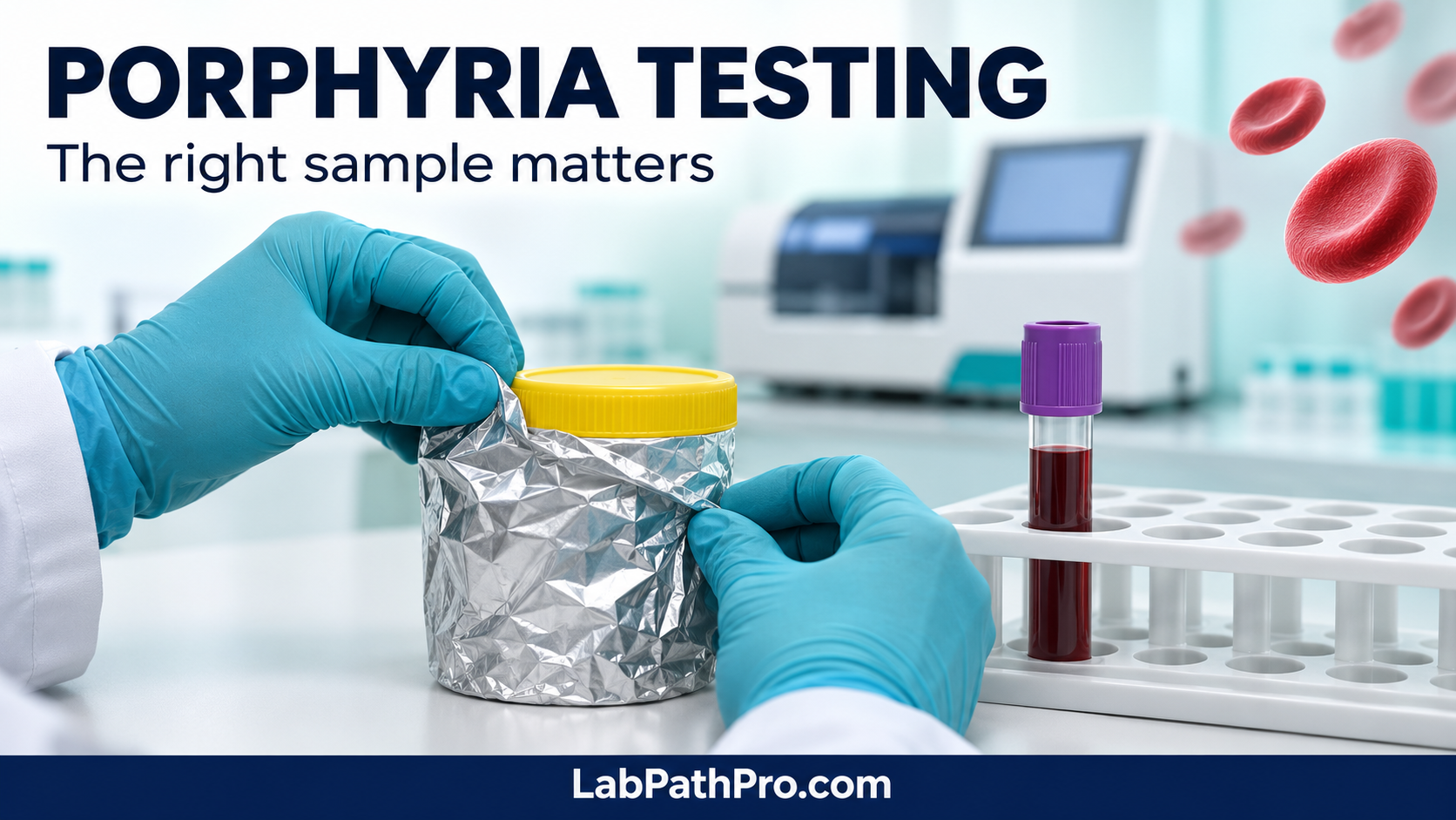
Collect specimens under normal lighting, then protect them from light immediately using an opaque container or foil. Do not leave samples exposed while awaiting transport. For acute investigations, fresh random urine without preservative is recommended; follow the receiving laboratory’s instructions for all specimen types. 4
Arrange prompt transport and confirm refrigeration or freezing requirements with the testing service. Avoid repeated freeze–thaw cycles. Supply symptom onset, current features, relevant treatment and suspected subtype. If an urgent attack is suspected, telephone the laboratory so the specimen and result can be prioritised. 4
Avoid common interpretation traps
- Red urine is a clue, not a diagnosis. Appearance cannot identify a porphyria or replace biochemical measurement. 5
- Mildly raised total porphyrins are non-specific. Secondary increases can occur with other illnesses, including hepatobiliary disease. 5
- A negative test answers only its intended question. Negative urinary PBG does not exclude all cutaneous porphyrias. 56
- PCT is not simply adult CEP. It has a different biochemical basis and may be associated with liver-related susceptibility factors. 8
Test your laboratory knowledge
Teaching scenario: an infant has red-stained nappies, blistering on exposed skin and anaemia. Would isolated urine PBG adequately investigate suspected CEP?
Reveal the learning point
No. Discuss a specialist cutaneous porphyria investigation, including porphyrin profiling and subsequent confirmation. Compare this with urgent investigation of a possible acute attack, where random urine PBG, ALA and creatinine address the immediate biochemical question.
Porphyria, especially CEP, is sometimes linked with vampire folklore because of photosensitivity and other physical features. The historical link is unproven. CEP requires porphyrin profiling; urinary PBG is central to investigating most acute hepatic porphyria attacks.
Further learning: explore our Clinical Chemistry resources and laboratory abbreviations.
Educational notice: This article supports Biomedical Science learning and does not replace clinical assessment or local laboratory procedures. Suspected acute porphyria with severe symptoms requires urgent medical assessment. People with porphyria need advice appropriate to their specific diagnosis.
References and further reading
- Erwin A, Balwani M, Desnick RJ. Congenital erythropoietic porphyria. GeneReviews. Read GeneReviews.
- International Porphyria Network. Congenital erythropoietic porphyria. Specialist guidance.
- Aarsand AK, To-Figueras J, Whatley S, Sandberg S, Schmitt C. Practical recommendations for biochemical and genetic diagnosis of the porphyrias. Liver Int. 2025;45(3):e16012. doi:10.1111/liv.16012.
- International Porphyria Network. Frequently asked questions on acute hepatic porphyria diagnosis. Diagnostic FAQs.
- International Porphyria Network. Laboratory diagnosis. Laboratory guidance.
- International Porphyria Network. Erythropoietic protoporphyria and X-linked erythropoietic protoporphyria. EPP guidance.
- Brunning A. Periodic Graphics: Porphyria and the vampirism myth. Chemical & Engineering News. 24 October 2024. Chemistry explanation.
- International Porphyria Network. Porphyria cutanea tarda. PCT guidance.