
Acute Kidney Injury: Creatinine, GFR and AKI Staging Explained for Biomedical Scientists
An unexpected rise in creatinine can be an early laboratory indication of acute kidney injury (AKI). For Biomedical Scientists, recognising the significance of that change requires more than checking whether a result exceeds its reference interval. It involves understanding the assay, comparing previous results, identifying potential errors and communicating clinically significant findings promptly.
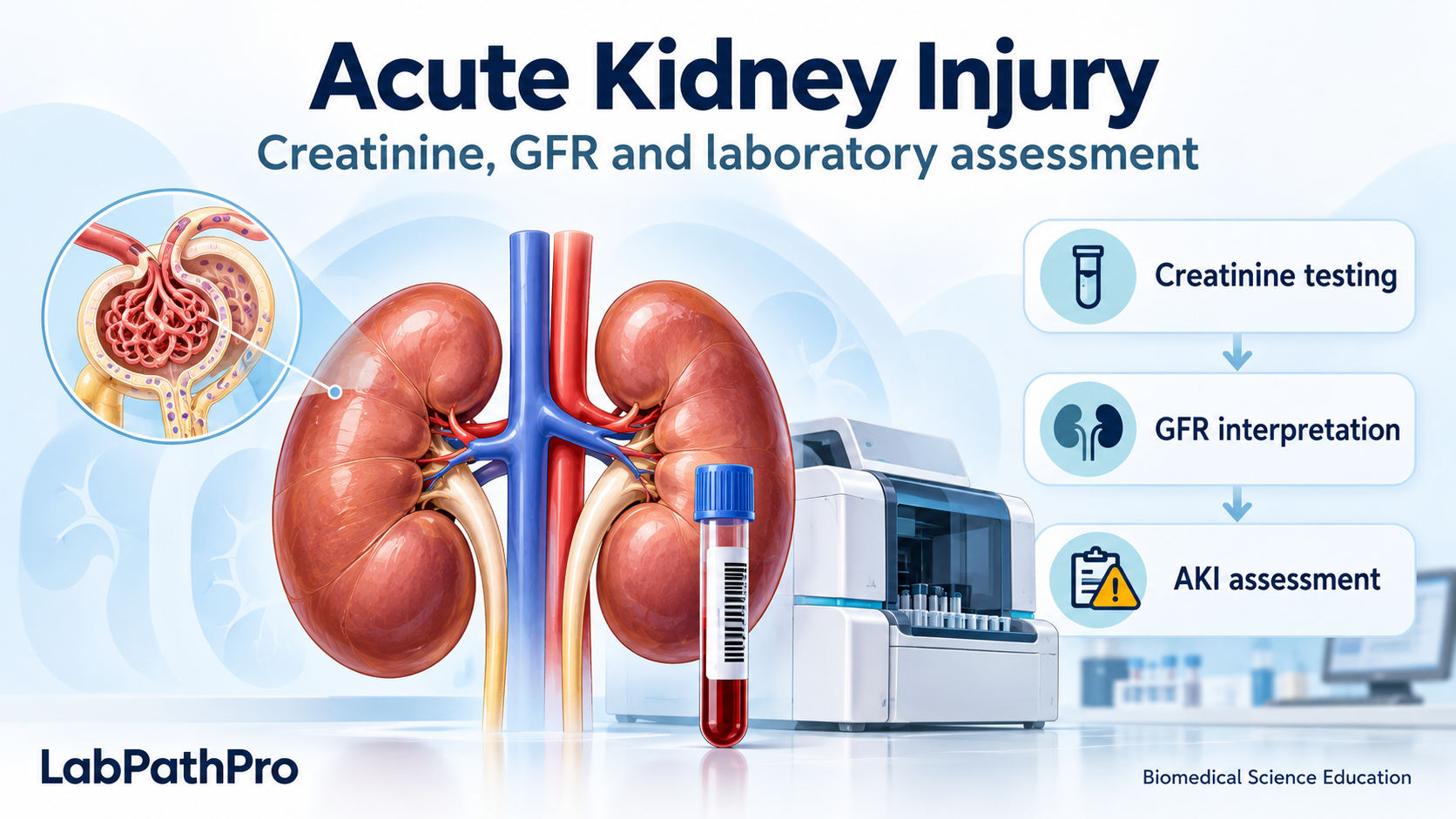
This article draws on the review by Chen and colleagues (2026), which explores AKI definitions, mechanisms and emerging diagnostic approaches 1. Established NICE and KDIGO guidance supports the practical laboratory sections 2–3–4. The focus is adult practice; paediatric assessment requires age-appropriate guidance.
What is acute kidney injury?
AKI is a rapid deterioration in kidney function. Reduced filtration can lead to the accumulation of waste products and disturbances in fluid, electrolyte and acid–base balance. AKI ranges from relatively small changes in creatinine to severe dysfunction requiring kidney replacement therapy.
The term does not mean that every patient has permanent kidney damage. Recovery is possible, although an episode increases the importance of subsequent monitoring for persistent dysfunction and chronic kidney disease (CKD).
Three broad categories help explain its causes:
| Category | What happens? | Examples |
|---|---|---|
| Pre-renal | Kidney perfusion falls, initially without necessarily causing structural injury | Hypovolaemia, haemorrhage or reduced cardiac output |
| Intrinsic renal | Disease or injury affects kidney tissue | Tubular injury, glomerulonephritis or acute interstitial nephritis |
| Post-renal | Obstruction prevents normal urine drainage | Bilateral urinary tract obstruction or obstruction of a solitary functioning kidney |
These categories can overlap. Sepsis-associated AKI, for example, involves complex circulatory, inflammatory and cellular processes. Chen and colleagues describe mitochondrial dysfunction, inflammation and regulated cell death as important mechanisms 1. For the laboratory, the practical distinction is that reduced filtration and cellular injury are related processes, but routine creatinine measures do not directly quantify tubular damage.
Why do we measure creatinine?
Creatinine is produced from creatine and phosphocreatine metabolism, mainly in skeletal muscle. It is cleared predominantly through glomerular filtration, with a smaller contribution from tubular secretion. When filtration falls, its concentration generally rises.
However, creatinine is an indirect marker of kidney function. Its concentration also reflects muscle mass, production rate, diet, distribution volume and some medicines. A patient with low muscle mass may have significant impairment despite a relatively modest creatinine concentration. Fluid accumulation can dilute creatinine and make deterioration less apparent.
Creatinine also takes time to accumulate after an abrupt fall in filtration. A reassuring initial result therefore does not exclude evolving AKI. Serial measurements and clinical information are essential.
A reference interval is not an AKI decision limit
Suppose creatinine rises from 50 to 80 µmol/L within 48 hours. The increase is 30 µmol/L and the ratio is 1.6. Even if 80 µmol/L falls within the laboratory’s adult reference interval, the change meets biochemical AKI criteria.
Conversely, a persistently raised result does not establish AKI without evidence of an acute change. The patient may have CKD, and AKI can also occur on top of CKD.
How is creatinine measured in the laboratory?
Jaffé methods
The Jaffé reaction uses the colour produced when creatinine reacts with alkaline picrate. Automated methods commonly measure the rate of colour development. Compensated methods apply corrections for some non-creatinine contributions.
The reaction is not completely specific. Depending on the method, glucose, ketones, proteins and certain cephalosporins can affect the result; bilirubin may cause negative interference. The direction and size of interference depend on the analytical system. Primary assay studies demonstrate that cephalosporin effects vary between methods 6.
Enzymatic methods
Enzymatic assays use a sequence of reactions to generate a measurable signal related to creatinine concentration. Many systems use a hydrogen peroxide-dependent colour reaction, although assay designs differ.
These methods generally offer greater specificity, but they are not free from interference. Biomedical Scientists should consult the manufacturer’s instructions and local validation data rather than assume that an enzymatic result is always correct.
NICE recommends specific creatinine assays, such as enzymatic methods, with calibration aligned to isotope dilution mass spectrometry (IDMS) 3. IDMS is a reference measurement approach: routine laboratories generally use assays whose calibration is traceable to it, rather than measuring every patient specimen by IDMS.
Why analytical quality matters for AKI
AKI detection depends on differences between results. Analytical bias, imprecision or a method change can affect whether an alert threshold is crossed. Internal quality control, external quality assessment and verification of reagent or calibration changes therefore support clinical interpretation, as well as analytical performance.
When investigating an unexpected rise, consider specimen identity, collection time, sample integrity, assay flags, quality-control performance, historical results and possible interference. Repeating the same sample can assess reproducibility, but it cannot exclude misidentification or contamination at collection.
GFR, eGFR and creatinine clearance: what is the difference?
Glomerular filtration rate (GFR) describes the volume of plasma filtered by the glomeruli per unit time. Laboratory reports usually provide an estimated GFR (eGFR) calculated from creatinine and demographic variables, using the locally adopted validated equation. It is commonly reported in mL/min/1.73 m², normalised to a standard body surface area.
| Measure | How it is obtained | Main limitation or distinction |
|---|---|---|
| Creatinine-based eGFR | An equation using serum creatinine and relevant demographic information | Assumes approximately stable creatinine kinetics; unreliable during rapid change |
| Measured GFR | Clearance of an exogenous filtration marker, such as iohexol, using a defined protocol | Requires a specialist procedure and is not usually part of urgent AKI testing |
| Creatinine clearance | Urine creatinine × urine flow rate ÷ plasma creatinine, with consistent units | Depends on timed urine collection and may overestimate GFR because creatinine is secreted by tubules |
Why eGFR is unreliable in AKI
During rapidly developing AKI, creatinine has not reached a new steady state. The calculated eGFR may therefore overestimate current filtration while creatinine is rising. During recovery, creatinine can remain elevated after filtration improves, causing eGFR to underestimate current function.
NICE identifies AKI as a situation in which creatinine-based eGFR is less reliable 3. Adult AKI staging uses changes in creatinine and urine output, rather than CKD eGFR categories. A single low eGFR during an acute illness also cannot establish CKD, which requires evidence of chronicity.
How is AKI detected?
For adults, NICE describes AKI detection using any of the following 2:
- A creatinine increase of at least 26 µmol/L within 48 hours.
- A creatinine increase of at least 50%, known or presumed to have occurred within the preceding 7 days.
- Urine output below 0.5 mL/kg/hour for more than 6 hours.
KDIGO expresses the absolute creatinine threshold as 0.3 mg/dL, equivalent to approximately 26.5 µmol/L, and uses a urine-output duration of at least 6 hours 4. Apply the exact thresholds in the validated local algorithm and SOP.
The time window matters: the relative creatinine criterion applies within 7 days, while the absolute increment applies within 48 hours. The review’s abbreviated definition places the relative rise within 48 hours; the criteria above follow the original guidance.
AKI staging: understanding severity
The established KDIGO framework is shown below 4. Use the highest stage met by either creatinine or urine-output criteria. Creatinine-based stages should be interpreted after establishing that the change is acute.
| Stage | Serum creatinine criteria | Urine-output criteria |
|---|---|---|
| 1 | 1.5 to less than 2 times baseline, or an increase of at least 26.5 µmol/L within 48 hours | Below 0.5 mL/kg/hour for 6 to less than 12 hours |
| 2 | 2 to less than 3 times baseline | Below 0.5 mL/kg/hour for at least 12 hours |
| 3 | At least 3 times baseline; or an acute increase to at least 353.6 µmol/L; or initiation of kidney replacement therapy | Below 0.3 mL/kg/hour for at least 24 hours, or anuria for at least 12 hours |
Anuria means absence of urine production. A chronically stable creatinine above 353.6 µmol/L does not, by itself, establish stage 3 AKI. The paediatric framework also includes an eGFR criterion for stage 3, which is outside this adult-focused table.
Worked examples
Assume an appropriate baseline, an acute rise within the required timeframe and no higher-stage urine-output criterion:
| Baseline → current creatinine | Calculation | Creatinine-based stage |
|---|---|---|
| 80 → 130 µmol/L within 48 hours | 130 ÷ 80 = 1.625; increase = 50 µmol/L | Stage 1 |
| 80 → 180 µmol/L within 7 days | 180 ÷ 80 = 2.25 | Stage 2 |
| 80 → 250 µmol/L within 7 days | 250 ÷ 80 = 3.125 | Stage 3 |
A patient meeting stage 1 creatinine criteria but stage 3 urine-output criteria is clinically classified as stage 3. The laboratory may not receive urine-output data, so its automated warning cannot always represent the complete clinical stage.
What does a laboratory AKI warning mean?
The NHS England algorithm compares current creatinine with historical results. Reference values include the lowest creatinine in the preceding 7 days and the median of results from 8–365 days previously; it also considers the absolute increase over 48 hours 5. The baseline is therefore not necessarily the immediately preceding result.
The resulting warning identifies a pattern requiring clinical assessment. It does not determine the cause of AKI or replace review of urine output and the patient’s condition. Missing historical results, incomplete records or an inappropriate baseline can affect detection. Absence of an alert does not exclude AKI.
For Biomedical Scientists, a practical workflow is:
- Validate the specimen and result: check identity, integrity, analyser flags and analytical acceptability.
- Review the trend: inspect previous creatinine values and sampling intervals.
- Check the warning: understand which comparison generated it and identify any discrepancy.
- Review associated results: particularly potassium and markers of acid–base disturbance.
- Escalate according to the SOP: communicate significant findings, document the recipient and time, and follow the escalation pathway if contact fails.
Investigating an unexpected result should occur alongside timely escalation where urgent risk is suspected. An AKI warning and a critical potassium result may have separate reporting requirements.
Which other tests support AKI assessment?
| Test | Laboratory contribution |
|---|---|
| Potassium | Detects hyperkalaemia, a potentially life-threatening complication; assess haemolysis and other causes of spurious elevation |
| Urea | Supports assessment of nitrogenous waste accumulation, but is also affected by catabolism, gastrointestinal bleeding and other factors |
| Bicarbonate or blood gas | Helps identify acid–base disturbance, including metabolic acidosis |
| Urine dipstick and microscopy | Blood, protein and sediment findings may guide investigation of the underlying cause |
| Creatine kinase | Supports investigation when rhabdomyolysis is suspected |
| Urine ACR or PCR | Quantifies albumin or protein when clinically indicated; interpretation needs the acute clinical context |
Testing follows clinical indications and local pathways. No single routine test identifies every cause of AKI.
Beyond creatinine: emerging biomarkers
Chen and colleagues discuss NGAL, KIM-1 and the combined urinary markers TIMP-2 and IGFBP7, alongside molecular diagnostics and newer approaches to GFR measurement 1. These approaches aim to identify tubular injury or cellular stress earlier than conventional changes in filtration markers.
The distinction is useful for portfolio learning: creatinine reflects filtration indirectly, while injury and stress biomarkers may provide different biological information. Their clinical roles depend on the assay, patient population and validated pathway. They should not be presented as universally available replacements for routine creatinine testing.
Applying this learning to an IBMS portfolio
A strong portfolio contribution connects scientific knowledge with your own laboratory practice. Useful evidence could include:
- An anonymised case review: show the creatinine trend, calculate the relevant increase and explain the warning or stage.
- An assay comparison: describe your laboratory’s method, calibration traceability and relevant interferences, supported by the SOP and manufacturer’s instructions.
- A result-validation reflection: explain the checks you performed, the limits of those checks and the significance of associated results.
- Observed communication: document a supervised escalation, including the information conveyed and how communication was recorded.
- A small audit: assess an agreed aspect of AKI reporting, such as alert consistency or documented communication, with appropriate approval.
Reading this article can support knowledge development; practical competence requires evidence of what you personally did. Map evidence only to the outcomes and HCPC Standards of Proficiency actually demonstrated, using the current portfolio and your trainer’s guidance. Remove patient identifiers and explain any screenshots or diagrams with clear legends.
Reflection prompts include: Why could AKI occur with creatinine inside the reference interval? How could assay interference affect an alert? Why is eGFR unreliable during rapid change? What would you do if an AKI warning accompanied a critical potassium result?
The laboratory’s contribution is accurate measurement, informed validation and effective communication. Understanding how a creatinine result becomes an AKI warning helps Biomedical Scientists connect analytical quality with patient care.
References
- Chen J, Xu X, Cai Y, Zhang Q, Wang X, Zhang D. (2026). Acute kidney injury over the past decade: from definition evolution to pathogenesis insights and innovative therapeutic strategies. Cellular and Molecular Life Sciences, 83, 156. https://doi.org/10.1007/s00018-026-06104-5
- NICE. Acute kidney injury: prevention, detection and management (NG148). https://www.nice.org.uk/guidance/ng148/chapter/Recommendations
- NICE. Chronic kidney disease: assessment and management (NG203). https://www.nice.org.uk/guidance/ng203/chapter/Recommendations
- KDIGO. (2012). Clinical Practice Guideline for Acute Kidney Injury. https://kdigo.org/wp-content/uploads/2016/10/KDIGO-2012-AKI-Guideline-English.pdf
- NHS England. Acute Kidney Injury Algorithm. https://www.england.nhs.uk/akiprogramme/aki-algorithm/ See also the primary implementation study: Consistency of alerts generated by, and implementation of, the NHS England acute kidney injury detection algorithm in English laboratories. https://doi.org/10.1007/s40620-024-02030-6
- Analytical interference of drugs in clinical chemistry: II — The interference of three cephalosporins with the determination of serum creatinine concentration by the Jaffé reaction. https://pubmed.ncbi.nlm.nih.gov/4092352/
Guidance checked: 17 October 2026. KDIGO’s website describes the 2026 AKI/AKD guideline as undergoing preparation for publication following public review. This article uses the published 2012 staging framework alongside current NICE guidance. Review the article when the final update is published: KDIGO AKI/AKD guideline update.
Continue your biomedical science learning
Explore the LabPathPro Learning Centre and Clinical Chemistry resources. Connect your own learning with IBMS Registration Portfolio guidance, our evidence-based practice guide and CPD guidance.
Educational use only: this article supports learning and professional discussion. It does not replace current national guidance, manufacturer instructions, local SOPs, formal risk assessment, clinical advice or professional judgement. Please read the full LabPathPro Disclaimer and Professional Notice.