
Breast Cancer Diagnosis: The Role of Biochemistry and the Medical Laboratory
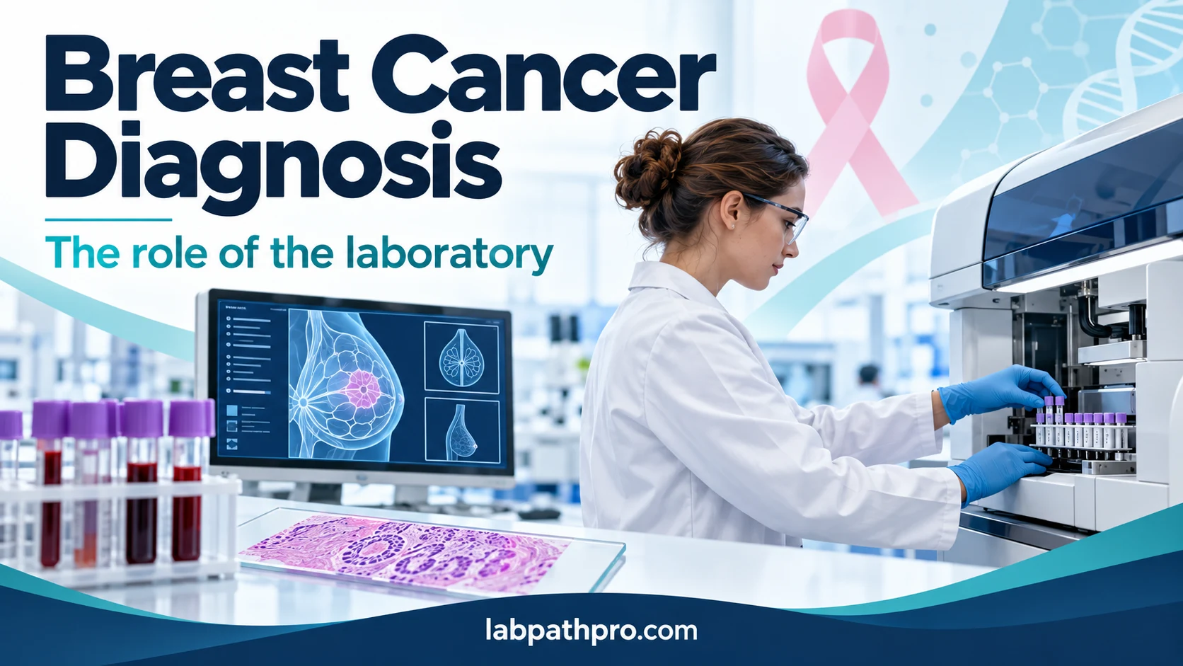
Breast cancer diagnosis depends on several pieces of evidence coming together: clinical assessment, breast imaging and examination of tissue. Behind this pathway, medical laboratories also provide information that helps clinicians select treatment, assess organ function and recognise complications.
For biomedical scientists, understanding these different roles matters. A blood test can be essential to safe cancer care without being a test that diagnoses the cancer itself.
Breast cancer in the UK: understanding the figures
Around 59,000 women and 420 men are diagnosed with breast cancer each year in the UK, according to Cancer Research UK. These annual averages refer to invasive breast cancer and use data from 2019 and 2021–2022; they are not a count of diagnoses made in 2026. Approximately 1 in 7 women in the UK will develop breast cancer during their lifetime. Risk generally increases with age, although younger adults can also develop the disease. [1,2]
Breast cancer can affect people of different sexes and gender identities. Anyone who notices an unexplained breast or chest change should seek medical advice.
Changes that should be checked
Be familiar with how your breasts or chest normally look and feel. Changes worth discussing with a GP include:
- A new lump or swelling in the breast, chest or armpit.
- Dimpling, puckering or an orange-peel appearance of the skin.
- A new colour change, redness or swelling; redness may be less obvious on darker skin.
- A change in the shape or size of a breast.
- A nipple newly turning inwards.
- A persistent rash, scaling or crusting involving the nipple.
- Unexpected nipple discharge, particularly blood-stained discharge. [3]
Most breast changes are not cancer, but a new or unusual change needs assessment. Do not wait for your next screening appointment or rely on a normal blood test for reassurance. Earlier diagnosis generally offers a better opportunity for successful treatment. [3]
How is breast cancer diagnosed?
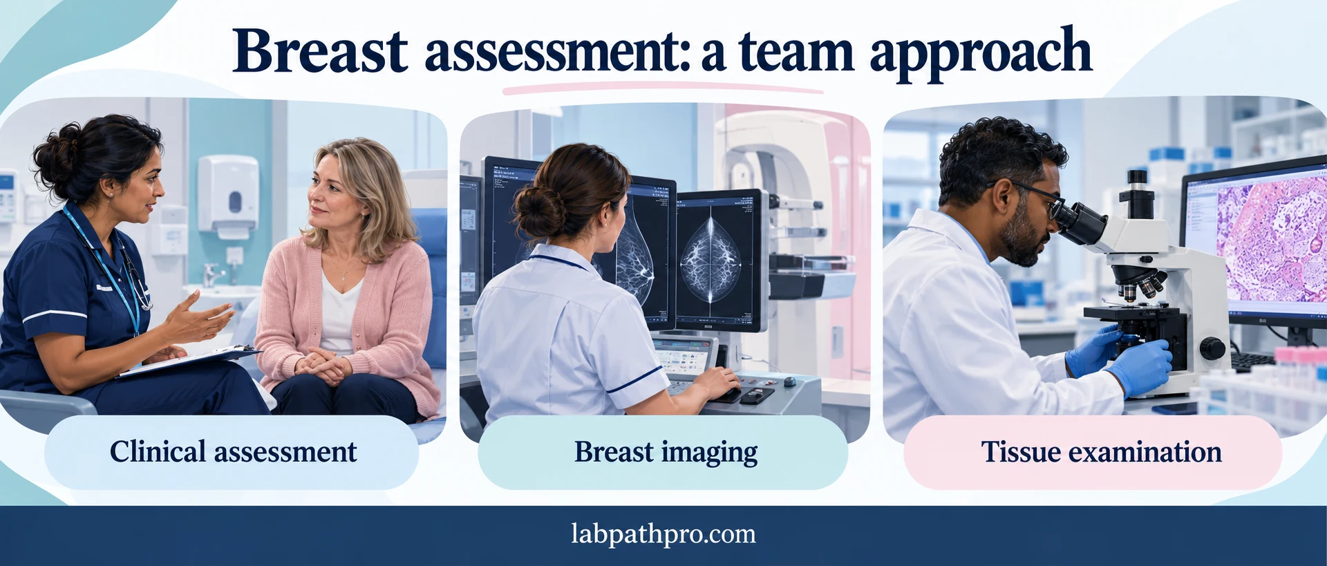
Specialist assessment commonly brings together three elements, often called triple assessment:
- Clinical assessment: a history and examination of the breasts or chest and nearby lymph nodes.
- Imaging: mammography and/or ultrasound, selected according to the person and clinical findings.
- Sampling: usually a core needle biopsy where indicated, allowing tissue to be examined in the laboratory. [4]
Not every person needs every investigation. A biopsy helps establish whether a suspicious abnormality is cancer and what kind it is. Further scans are selected when needed to assess the extent of disease; a CT, PET-CT or MRI scan is not automatically required for everyone. [4,5]
Screening and symptomatic assessment serve different purposes. Screening aims to find cancers before symptoms develop. A new symptom requires clinical assessment even after a recent normal screening result. [3,6]
Histopathology: establishing the diagnosis and tumour profile
In histopathology, tissue is processed into sections, stained and assessed microscopically. This helps the pathologist determine the tumour type and grade. Surgical specimens can provide further information, including margin and lymph node findings.
Additional tissue tests help guide treatment. NICE recommends assessing oestrogen receptor (ER), progesterone receptor (PR) and human epidermal growth factor receptor 2 (HER2) status in all invasive breast cancers at initial histopathological diagnosis. These markers describe characteristics of the tumour; they are not routine serum hormone measurements. [7]
ER and PR testing uses immunohistochemistry, which uses antibodies to demonstrate proteins in tissue. HER2 assessment may involve immunohistochemistry and, where appropriate, in situ hybridisation to investigate gene amplification. Results help the multidisciplinary team identify suitable endocrine or HER2-directed therapies. [7,8]
Although the word “hormone” appears in this pathway, measuring circulating oestrogen in a biochemistry sample does not establish whether a breast tumour is ER-positive.
Where does the biochemistry laboratory fit?
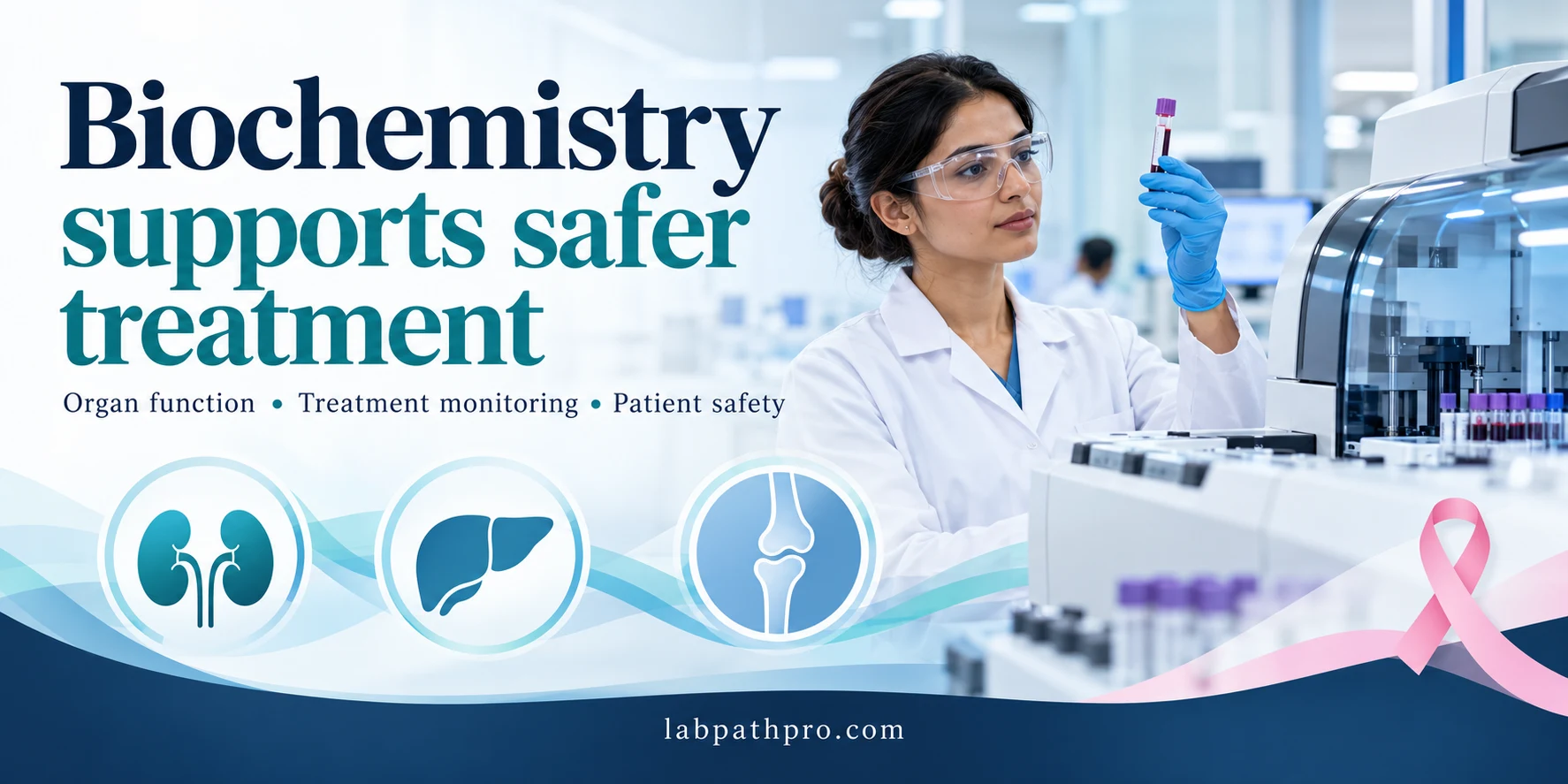
Biochemistry contributes mainly to assessment before treatment, monitoring during treatment and investigation of complications. The requested tests and frequency depend on the treatment regimen, symptoms, medical history and local oncology protocol. They are not a universal breast cancer blood-test panel. [4,9]
On smaller screens, swipe the table sideways to read all three columns.
| Test or group | What it contributes | Important limitation |
|---|---|---|
| Creatinine, urea, electrolytes and estimated glomerular filtration rate (eGFR) | Information about kidney function and electrolyte balance, supporting treatment safety and prescribing decisions | A normal kidney profile does not exclude breast cancer; drug dosing may require a regimen-specific renal assessment |
| Alanine aminotransferase (ALT), alkaline phosphatase (ALP), bilirubin and albumin | Information about liver-related abnormalities and the effects of illness or treatment | Abnormal results have many causes and cannot independently establish liver metastases |
| Calcium and bone profile | Assessment of calcium disturbances and support for investigation of bone-related complications | A raised calcium or ALP result does not, by itself, diagnose bone metastases |
| Magnesium and other targeted chemistry tests | Investigation of losses or disturbances during treatment, where clinically indicated | Testing should answer a clinical question rather than be requested indiscriminately |
These applications are consistent with NHS chemotherapy blood-testing information. Interpretation requires the clinical picture and other investigations. [9]
A full blood count is also important in cancer care, including assessment of anaemia, neutrophils and platelets. It is primarily a haematology investigation, rather than a biochemistry test. This distinction illustrates how laboratory departments work together. [10]
Can CA 15-3 diagnose breast cancer?
No. CA 15-3 should not be used to screen for breast cancer or to exclude it.
CA 15-3 is a circulating tumour-associated marker measured by an immunoassay. It may help monitor selected patients with established disease, particularly advanced breast cancer, when the oncology team considers it useful. However:
- Some breast cancers do not produce an elevated result.
- Non-cancerous conditions and other cancers can increase the concentration.
- Early treatment-related changes may not reflect disease progression.
- A result must be interpreted alongside symptoms, examination and imaging. [11,12]
The useful question is often whether a meaningful trend supports the other evidence, rather than whether a single value sits above a reference limit. Serial measurements should, where possible, use a consistent laboratory method. A reference interval is method-dependent and is not a diagnostic threshold for breast cancer.
Routine tumour-marker testing is not a substitute for the agreed follow-up plan after treatment for early breast cancer. A treatment change should not be based on CA 15-3 alone. [4,7]
A practical laboratory example
Consider a person receiving treatment for established breast cancer whose ALP concentration has risen. The result may prompt review, but it does not reveal its cause on its own. The clinical team considers symptoms, previous results, other liver-related tests, medicines and relevant imaging.
Similarly, a normal calcium concentration cannot rule out bone involvement. The laboratory contributes evidence to the investigation; it does not determine the diagnosis from one analyte.
For biomedical scientists, the practical priorities include correct patient identification, suitable specimens, recognition of sample problems, reliable quality control, timely reporting and escalation of critical results according to local procedures. Unexpected findings should prompt appropriate checks and discussion with senior laboratory staff or the clinical team.
Liquid biopsy: a different kind of blood test
A liquid biopsy can examine circulating tumour DNA (ctDNA): small fragments of tumour-derived DNA present in plasma. This is different from a routine chemistry profile or a CA 15-3 immunoassay.
In NHS England, ctDNA testing is already commissioned for specified breast cancer indications, including ESR1 testing linked to elacestrant eligibility and selected PIK3CA/AKT1/PTEN testing to support targeted treatment decisions. These are specialist genomic investigations for eligible patients, rather than general screening tests. [13,14]
ESR1 encodes the oestrogen receptor. Acquired changes in this gene can contribute to resistance to certain endocrine treatments. Identifying an actionable change can therefore help select a subsequent treatment. NICE specifies eligibility criteria for elacestrant, including progression after at least 12 months of endocrine treatment combined with a CDK4/6 inhibitor in the relevant population. [15]
A negative plasma result may be uninformative when too little tumour DNA is circulating. It does not prove that cancer is absent. Specialist teams determine whether further testing is appropriate. Availability and pathways should be checked within each UK nation and local genomic service. [14]
What should readers take away?
Breast cancer diagnosis is a collaborative process. Clinical assessment and imaging identify abnormalities; tissue examination establishes the diagnosis and characterises the tumour. Biochemistry supports safer treatment and investigation of complications, while specialist genomics can identify treatment-relevant changes in selected patients.
If you notice a new breast, chest or nipple change, contact your GP. Normal routine blood tests or a normal CA 15-3 result cannot exclude breast cancer.
This article provides education and does not replace individual assessment or advice from a healthcare professional.
References and further reading
- Cancer Research UK. Breast cancer incidence (invasive) statistics. Annual averages: 2019 and 2021–2022.
- Cancer Research UK. Breast cancer: overview and lifetime risk.
- NHS. Symptoms of breast cancer in women.
- Cancer Research UK. Tests for breast cancer.
- NHS. Tests and next steps for breast cancer in women.
- NHS. Breast screening.
- NICE. NG101: Early and locally advanced breast cancer—recommendations. See tumour profiling and follow-up sections.
- NICE. NG101: Full guideline, including HER2 testing methods.
- Leeds Teaching Hospitals NHS Trust. Blood testing for patients receiving chemotherapy.
- Macmillan Cancer Support. Blood tests.
- Lab Tests Online UK, Association for Laboratory Medicine. CA 15-3 test.
- Canadian Cancer Society. Cancer antigen 15-3 test.
- NHS England. Implementation of circulating tumour DNA testing in the NHS. April 2026.
- South East Genomics. Changes to the Cancer Genomic Test Directory.
- NICE. TA1036: Elacestrant for treating ER-positive HER2-negative advanced breast cancer with an ESR1 mutation after endocrine treatment.
All sources accessed 25 October 2026.